


8 October 2026
A transplanted heart may be being rejected before symptoms appear, and an electrocardiogram could help detect it early
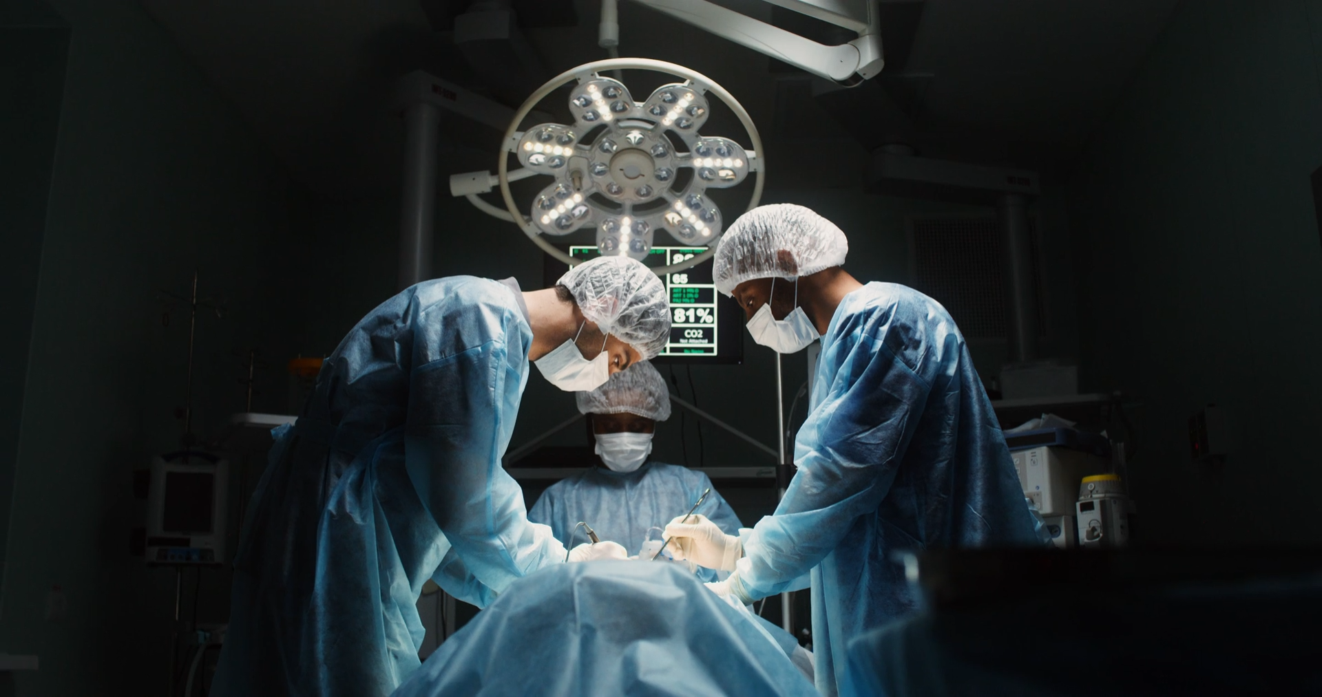
Receiving a new heart does not mean that the body immediately recognizes it as its own. For the immune system, that organ is a foreign body and, despite treatment with immunosuppressants, there is a risk that it will begin to attack it. The problem is that this rejection does not always give warning: it can develop without the patient noticing any symptoms.
Precisely for this reason, after a heart transplant, follow-up is not limited to checking how the person is doing. During the first months, frequent check-ups are performed, and one of the main tools to look for signs of rejection remains the endomyocardial biopsy, in which small fragments of the heart are obtained for microscopic analysis.
Now, researchers at NYU Langone Health have explored a much less invasive alternative: using artificial intelligence to analyze the information contained in an electrocardiogram and combine it with biomarkers present in the blood. The results obtained from this new technique indicate that this mixture of signals could help identify which patients have a very low probability of experiencing rejection and, thereby, avoid some unnecessary biopsies.
A body trying to defend itself from a heart it needs
Rejection arises from an inevitable paradox of transplantation. The immune system is designed to recognize what does not belong to the body and react against it. This ability is essential against numerous external agents, but it also means that cells from a donor heart can be identified as foreign.
Immunosuppressive drugs reduce this response and are part of the long-term treatment for transplanted individuals. Still, rejection can occur days, weeks, months, or even years after the operation. Moreover, in some cases, it does not generate recognizable symptoms during its early stages.
When symptoms do appear, they may include shortness of breath, fatigue, fever, palpitations, fluid retention, or unexpected weight gain, among other manifestations. But waiting for symptoms to appear is not a sufficient strategy to monitor a transplanted organ, which is why check-ups are also performed when the patient appears to be well.
Looking directly at the heart tissue
The endomyocardial biopsy allows searching in the tissue itself for signs of inflammation and other changes compatible with immune system attack. To obtain the sample, a catheter is usually inserted through a vein in the neck or groin until it reaches the heart, where small fragments are extracted and later examined in the laboratory.
The frequency of these biopsies is usually especially high during the first months after transplantation, when the risk of rejection is greatest, and decreases progressively over time.
Therefore, finding a reliable way to rule out rejection without having to reach the heart tissue would reduce part of this invasive monitoring. And this is where a much more common and simple test comes into play: the electrocardiogram.
Signals we do not see at first glance
The study, published on September 25 in JHLT Open, gathered 5,300 electrocardiograms corresponding to 2,357 adults who had received a heart transplant. The researchers trained artificial intelligence models to identify patterns associated with rejection and compared their results with those obtained through biopsies.
The electrocardiogram records, via electrodes placed on the skin, the electrical activity accompanying each heartbeat. For a healthcare professional, it provides information about the heart’s rhythm and electrical conduction, among other aspects. However, a digital record also contains a huge amount of data that an algorithm can analyze collectively to locate much subtler patterns.
The idea is not, therefore, that an obvious signal appears on the electrocardiogram indicating that the organ is being rejected. The model looks for combinations of features within the electrical signal that relate to what the heart tissue subsequently shows.
This is not the first time this approach has been explored. A previous study conducted with 1,427 transplanted patients had already shown that a deep learning model applied to the electrocardiogram can identify cases of acute, moderate, or severe cellular rejection. The novelty comes from adding another source of information.
The electrocardiogram does not work alone
The researchers developed a model that combined the heart’s electrical recordings with two blood biomarkers used to estimate the risk of rejection: one related to genetic activity associated with cellular rejection and another based on donor-derived DNA fragments circulating in the recipient’s blood.
To train it, data from patients treated between 2018 and 2024 were used, and each electrocardiogram was linked to a biopsy performed within a maximum period of one month. Cases were classified into two large groups: absence or mild rejection versus moderate or severe rejection, the latter more likely to require changes in immunosuppressive treatment.
After that training came the ultimate test: checking how the system worked in 38 additional heart transplant recipients.
In that small group, the combined model correctly identified 94% of patients who were not experiencing rejection. And this data is important for what it implies: we do not claim that the test diagnoses any rejection with 94% accuracy, but it does show its ability to recognize those who do not have it.
Combining different procedures that individually would not yield the same result is another benefit of this study. While the model based solely on blood analyses mistakenly flagged 19 patients as possible candidates for biopsy, by also incorporating electrocardiogram information, those procedures would have been avoided.
Fewer biopsies, but still necessary
The results show the value of combining different signals. Blood biomarkers provide information about molecular processes related to the transplanted organ, while the electrocardiogram captures its electrical activity. Analyzing both data sets together allows for a more precise assessment of rejection risk.
Still, the electrocardiogram is far from replacing the biopsy. The independent group used to test the model consisted of only 38 people, and it will be necessary to evaluate it in larger samples and at different transplant centers.
Caution is justified because many of these models have still been tested on small groups or based on retrospective data. A review published in 2026 on artificial intelligence applied to heart and lung transplants emphasizes precisely this problem and the need to confirm results in more patients and different centers before broadly transferring these models to clinical practice.
For now, the biopsy remains a fundamental tool for monitoring rejection. The possibility raised by this work is to use data obtained in a much less invasive way beforehand to better identify which patients really need to undergo it.

