


22 September 2026
What happens when millions of human neurons develop inside an animal's brain?
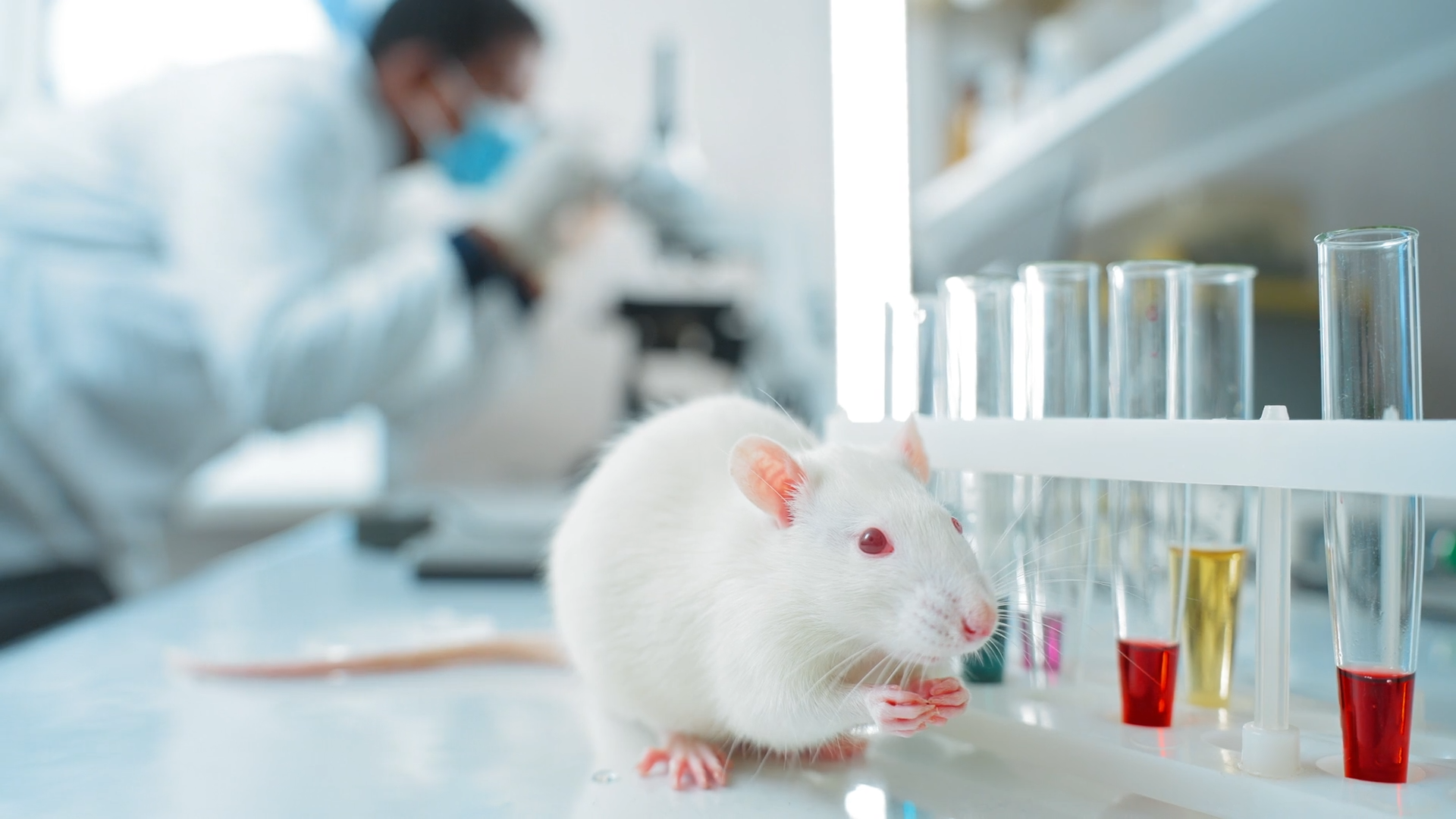
The human brain remains one of the most difficult organs to study while it is still functioning. Samples obtained after surgery or post-mortem allow observation of its structure; cell cultures show what happens at the microscopic scale; and organoids reproduce part of its development in three dimensions. But none of these models alone achieve something fundamental: tracking human neurons as they grow, form circuits, and respond within a living organism.
A team from Stanford University has just approached this scenario in an unusual way. The researchers implanted human cerebral cortex organoids into genetically modified mice whose own cortex and hippocampus largely failed to develop normally. Three months later, the tissue from the graft accounted for about 92% of the animals’ combined cortical volume.
But it did not just occupy space. The human cells matured, generated different neuronal types, extended axons to other regions of the nervous system, and showed organized electrical activity. The work, published on September 16 in Nature, opens a different way to study what happens when human brain tissue stops growing isolated in a dish and becomes part of another living circuit.
Making room before transplanting
Transplanting brain organoids into rodents is not a completely new idea. Other studies had already shown that these small three-dimensional structures, generated from stem cells, survive in the rodent brain and establish connections with the surrounding tissue. The problem was space.
The host animal’s neurons soon begin to form their own circuits and compete with the graft. Moreover, human cells develop much more slowly than those of a rodent. The result so far was tissue limited in size and forced to integrate into an architecture that was already underway.
The team led by Sergiu Pașca changed that starting condition. Through a genetic modification, they markedly reduced glutamatergic neurons in the neocortex and hippocampus. As a result, these mice developed a brain approximately 50% smaller than that of animals without this modification.
Shortly after birth, they received the human cortical organoids. Having space that would normally have been occupied by the mouse’s own cortex, the grafts grew much more extensively: between the second and third month their volume increased about 4.7 times.
Being inside the brain is not enough: it must be verified if the tissue works
One thing was to get the human tissue to grow inside the mouse brain and quite another to verify if it really integrated and functioned as part of that nervous system.
The researchers observed that the human neurons did not remain isolated. They extended connections to other brain regions and showed activity typical of developing neuronal circuits, a sign that the graft was not simply surviving.
Additionally, some types of neurons especially difficult to study in the lab appeared. Among them were cells with characteristics similar to Von Economo neurons, present in certain areas of the human brain and related to complex functions such as social processing.
The transplanted tissue was still far from reproducing a complete human cortex, but the experiment showed to what extent these cells were capable of developing and integrating into the animal’s nervous system.
A test with oxygen deprivation
The usefulness of the new model became especially clear when the researchers wanted to check how the human tissue reacted to severe oxygen deprivation.
This type of injury usually has significant consequences in the human brain, but it is difficult to study in mice because they are more resistant than us to its effects. The presence of the graft allowed observing a different response.
After keeping the animals for five hours with very low oxygen levels, the human tissue showed clear signs of damage that barely appeared in the mouse’s own brain cells. Afterwards, the researchers also detected changes in the animals’ coordination.
The experiment thus demonstrated one of the advantages of this model: it allows studying how human brain tissue responds to injury within a living organism, something that until now was much harder to reproduce in the laboratory.
From a patient’s organoid to a living circuit
One of the possibilities this model opens is creating organoids from cells of specific patients. By preserving part of their genetic characteristics, researchers can observe how that brain tissue develops within a living environment and what differences appear compared to normal development.
Until now, much of this work has been done in laboratory dishes, where cells grow without being part of a complete nervous system. Integrating them into a living brain would allow following their evolution for longer, verifying how they establish connections with other areas, and also studying their response to different treatments.
This step is especially interesting in brain developmental disorders that mice reproduce with difficulty. Their brains mature much faster and do not reflect all the processes typical of human neurons. Having tissue from patients integrated into a living organism brings the laboratory closer to conditions that until now were very difficult to reproduce.
Are these mice with a human brain?
The magnitude of the graft leads to an inevitable question: are these mice with a human brain? and the answer is no. Much of their brain and nervous system remains that of the animal itself, and the transplanted tissue reproduces only some aspects of a developing human cortex. The study also does not show that they have acquired human cognitive abilities.
However, the experiment does raise ethical questions about how far the integration of human cells into other species should go. Pașca has noted that he does not consider routinely transferring this procedure to species closer to humans, such as primates, where such integration would raise different concerns.
What changes with this work is the context in which organoids can be studied: human tissue has grown, established connections, and responded to injury within a living organism. This allows observing human brain processes that until now were very difficult to reproduce outside the laboratory.

